You will get CT Bone Segmentation and 3D Reconstruction for Medical AI
Project details
Radiologist with clinical CT experience performing high-accuracy medical image segmentation for AI datasets and research.
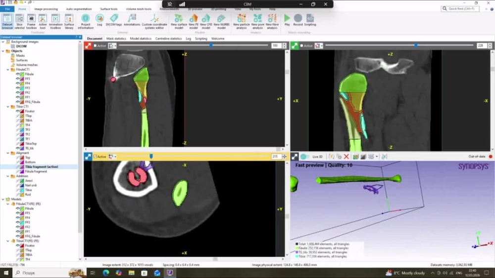
I provide professional medical image segmentation from CT datasets with a focus on bone structures, implants, and fracture fragments. As a radiologist experienced in medical imaging, I create accurate segmentation masks and high-quality 3D reconstructions suitable for orthopedic analysis, surgical planning, and AI training datasets. My workflow ensures precise anatomical labeling and clean models ready for research, machine learning, and medical visualization.
I provide professional medical image segmentation from CT datasets with a focus on bone structures, implants, and fracture fragments. As a radiologist experienced in medical imaging, I create accurate segmentation masks and high-quality 3D reconstructions suitable for orthopedic analysis, surgical planning, and AI training datasets. My workflow ensures precise anatomical labeling and clean models ready for research, machine learning, and medical visualization.
Machine Learning Tools
OpenCVWhat's included
| Service Tiers |
Starter
$50
|
Standard
$120
|
Advanced
$250
|
|---|---|---|---|
| Delivery Time | 3 days | 5 days | 7 days |
Number of Revisions | 1 | 2 | 3 |
Number of Model Variations | 1 | 2 | 3 |
Number of Scenarios | 1 | 1 | 1 |
Number of Graphs/Charts | 0 | 0 | 0 |
Model Validation/Testing | - | - | - |
Model Documentation | - | - | - |
Data Source Connectivity | - | - | - |
Source Code | - | - | - |
Optional add-ons
You can add these on the next page.
Fast Delivery
+$30 - $80
Additional Revision
+$20Frequently asked questions
About Olha
Radiologist | CT Imaging | Medical Image Segmentation | 3D Reconstruct
Vinnytsya, Ukraine - 6:04 pm local time
I combine clinical radiology expertise with medical image segmentation and preparation of medical imaging datasets for AI/ML projects, delivering anatomically accurate annotations and clinically meaningful analysis suitable for training deep learning models and medical imaging systems.
Radiology Expertise
• CT interpretation (neuro CT, CT angiography, spine and musculoskeletal imaging)
• X-ray interpretation, including orthopedic trauma imaging
• Mammography interpretation and BI-RADS classification
• Emergency and routine diagnostic imaging
• Structured radiology reporting
Clinical Experience
• 5760+ CT studies interpreted (brain, CT angiography, spine and musculoskeletal system)
• 1080+ mammography studies interpreted and reported using BI-RADS classification
• 600+ mammography images annotated for medical imaging AI datasets
• Extensive experience in orthopedic trauma CT and X-ray imaging
AI & Medical Image Segmentation
• 3D segmentation of skeletal structures (bones) from CT scans
• Pixel-level annotation and segmentation mask creation for AI datasets
• Medical image labeling and dataset preparation for deep learning models
• CT post-processing including MPR, MIP and CT post-processing including MPR, MIP and 3D volume rendering (VR)
• Preparation of structured datasets for AI training workflows
Segmentation Experience
• 100+ CT segmentations of tibia and fibula bones
• Tibia fracture cases treated with intramedullary nail fixation
• Fracture cases with extramedullary plate fixation
• Creation of anatomically accurate bone segmentation masks for AI training datasets
• Experience working with orthopedic trauma CT imaging
Tools & Platforms
• Simpleware – advanced medical image segmentation
• 3D Slicer – segmentation and 3D reconstruction
• PACS systems – clinical imaging workflow
• DICOM viewers (RadiAnt and clinical viewers)
• RedBrick AI – medical image annotation platform
• Excel – dataset organization and tracking
• Clinical reporting systems
Data Formats
• DICOM
• NIfTI
• 3D models (STL)
Services I Provide
• Medical image segmentation from CT scans
• Orthopedic CT segmentation (tibia / fibula fractures)
• Medical image annotation for AI datasets
• Radiological interpretation of CT and X-ray studies
• Mammography analysis and BI-RADS classification
• Structured radiology reporting
• Medical imaging dataset preparation and validation for AI models
• DICOM medical image analysis and labeling
✔ Detail-oriented radiologist experienced in medical imaging analysis, AI dataset annotation, and medical image segmentation, delivering accurate and clinically meaningful results for AI and research projects.
Steps for completing your project
After purchasing the project, send requirements so Olha can start the project.
Delivery time starts when Olha receives requirements from you.
Olha works on your project following the steps below.
Revisions may occur after the delivery date.
Step 1
Client provides CT DICOM dataset and segmentation requirements.
Step 2
I perform medical image segmentation, refinement, and 3D reconstruction.